![]()
Skill:
• Analysis of electron micrographs to find the state of contraction of muscle fibres
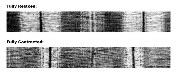
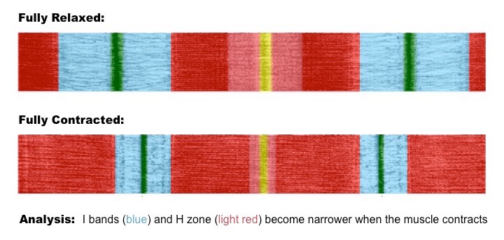
The arrangement of myofilaments within a sarcomere give a skeletal muscle fibre a striated appearance
- A sarcomere has a central darker region (A band) where actin and myosin filaments overlap
- A sarcomere has peripheral lighter regions (I bands) where actin is present, but not myosin
When muscle fibres contract, actin filaments slide along the myosin, reducing the length of the lighter I bands
- The movement of the actin filaments also reduces the width of the H zone, however the length of A bands do not change
Changes in Band Lengths with Sarcomere Shortening
Electron Micrographs of Muscle Fibres (Click for false colour imaging)