![]()
Understanding:
• Calcium ions and the proteins tropomyosin and troponin control muscle contractions
The process of muscular contraction occurs over a number of key steps, including:
- Depolarisation and calcium ion release
- Actin and myosin cross-bridge formation
- Sliding mechanism of actin and myosin filaments
- Sarcomere shortening (muscle contraction)
1. Depolarisation and Calcium Ion Release
- An action potential from a motor neuron triggers the release of acetylcholine into the motor end plate
- Acetylcholine initiates depolarisation within the sarcolemma, which is spread through the muscle fibre via T tubules
- Depolarisation causes the sarcoplasmic reticulum to release stores of calcium ions (Ca2+)
- Calcium ions play a pivotal role in initiating muscular contractions
Muscle Innervation
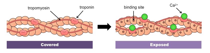
2. Actin and Myosin Cross-Bridge Formation
- On actin, the binding sites for the myosin heads are covered by a blocking complex (troponin and tropomyosin)
- Calcium ions bind to troponin and reconfigure the complex, exposing the binding sites for the myosin heads
- The myosin heads then form a cross-bridge with the actin filaments
The Role of Calcium in Cross-Bridge Formation
![]()
Understanding:
• The contraction of the skeletal muscle is achieved by the sliding of actin and myosin filaments
• ATP hydrolysis and cross bridge formation are necessary for the filaments to slide
3. Sliding Mechanism of Actin and Myosin
- ATP binds to the myosin head, breaking the cross-bridge between actin and myosin
- ATP hydrolysis causes the myosin heads to change position and swivel, moving them towards the next actin binding site
- The myosin heads bind to the new actin sites and return to their original conformation
- This reorientation drags the actin along the myosin in a sliding mechanism
- The myosin heads move the actin filaments in a similar fashion to the way in which an oar propels a row boat
Sliding Filaments Mechanism
4. Sarcomere Shortening
- The repeated reorientation of the myosin heads drags the actin filaments along the length of the myosin
- As actin filaments are anchored to Z lines, the dragging of actin pulls the Z lines closer together, shortening the sarcomere
- As the individual sarcomeres become shorter in length, the muscle fibres as a whole contracts
Diagrams of Sarcomere Shortening

Summary of Muscle Contractions
- Action potential in a motor neuron triggers the release of Ca2+ ions from the sarcoplasmic reticulum
- Calcium ions bind to troponin (on actin) and cause tropomyosin to move, exposing binding sites for the myosin heads
- The actin filaments and myosin heads form a cross-bridge that is broken by ATP
- ATP hydrolysis causes the myosin heads to swivel and change orientation
- Swiveled myosin heads bind to the actin filament before returning to their original conformation (releasing ADP + Pi)
- The repositioning of the myosin heads move the actin filaments towards the centre of the sarcomere
- The sliding of actin along myosin therefore shortens the sarcomere, causing muscle contraction
Muscle Contraction Summary
Please Note:
In this animation the myosin head is attached to actin when ATP hydrolysis causes it to swivel
In reality, the myosin head swivels when unattached and then returns to its original conformation following actin binding