
![]()
Skill:
• Identification of pneumocytes, capillary endothelium cells and blood cells in light micrographs and electron
micrographs of lung tissue
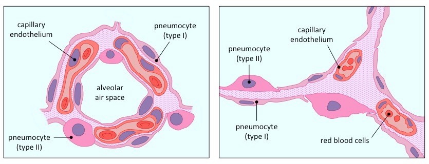
The inner surface of the alveolus is lined by a special type of alveolar cell called a pneumocyte
- Type I pneumocytes are very thin in order to mediate gas exchange with the bloodstream (via diffusion)
- Type II pneumocytes secrete a pulmonary surfactant in order to reduce the surface tension within the alveoli
Alveolar air spaces are surrounded by a dense network of capillaries, which transport respiratory gases to and from the lungs
- The capillaries are located close to the pneumocytes and are composed of a very thin, single-layer endothelium
- The capillaries transport oxygen within red blood cells, while white blood cells may extravasate into the lung tissue
Diagrammatic Representation of Lung Tissue
Light Micrograph of Lung Tissue (click to show / hide labels)


Electron Micrograph of Lung Tissue (click to show / hide labels)
