
![]()
Skill:
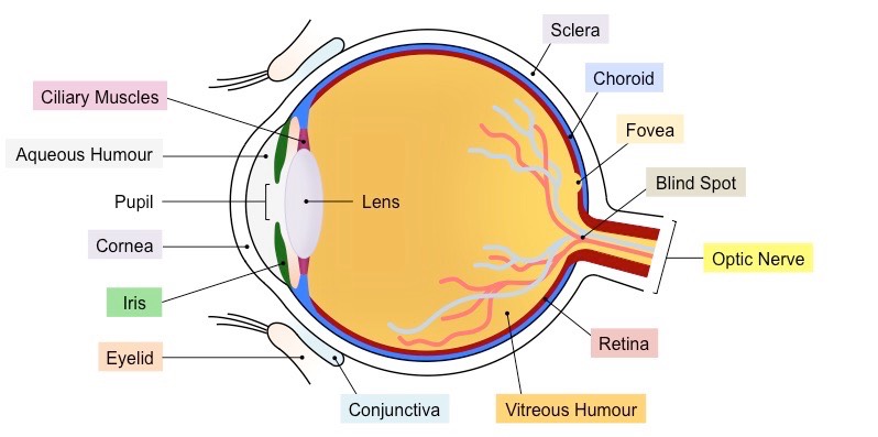
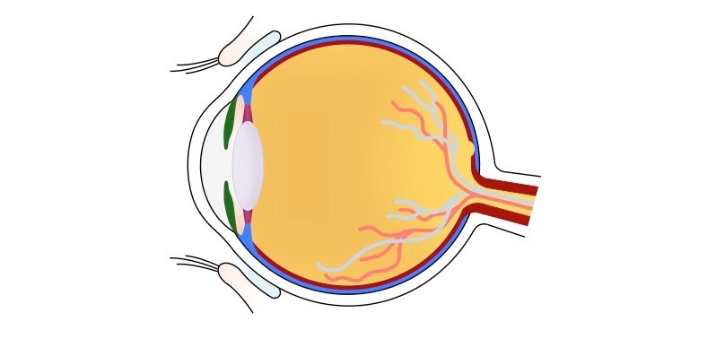
• Labelling a diagram of the structure of the human eye
The human eye is the sensory organ responsible for vision (sight perception)
- It consists of two fluid-filled cavities separated by a lens (anterior = aqueous humour, posterior = vitreous humour)
- The lens is attached to ciliary muscles, which can contract or relax to change the focus of the lens
- The amount of light that enters the eye via the pupil is controlled by the constriction and dilation of the iris
- The exposed portion of the eye is coated by a transparent layer called the cornea, which is lubricated by conjunctiva
- The internal surface of the eye is composed of three layers – the sclera (outer), choroid (middle) and retina (inner)
- The region of the retina responsible for sharpest vision (i.e. focal point) is the fovea centralis (or fovea for short)
- Nerve signals from the retina are sent via an optic nerve to the brain (no retina in this region creates a visual blind spot)
Diagram of the Human Eye
⇒ Click on the diagram to show / hide labels
![]()
Skill:
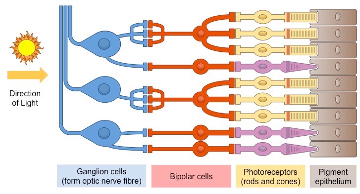
• Annotation of a diagram of the retina to show the cell types and the direction in which light moves
The retina is the light-sensitive layer of tissue that forms the innermost coat of the internal surface of the eye
- Two types of photoreceptors (rods and cones) convert light stimuli into electrical nerve impulses
- These nerve impulses are transmitted via bipolar cells to ganglion cells, whose fibres from the optic nerve tract
- The photoreceptors line the rear of the retina (adjacent to the choroid), meaning light passes through the other cell layers
Diagram of the Human Retina
![]()
Skill:
• Labelling a diagram of the structure of the human ear
The human ear is the sensory organ responsible for hearing (sound perception)
- The external part of the ear is called the pinna, whereas the internal part of the ear is divided into three sections
- The outer ear contains the auditory canal, which channel sound waves to the tympanic membrane (or eardrum)
- The middle ear contains three small bones called the ossicles, which transfer vibrations to the oval window
- The inner ear consists of the cochlea and semicircular canals, as well as a round window which dissipates vibrations
- The cochlear converts sound stimuli into electrical nerve impulses, which are transmitted via the auditory nerve to the brain
Diagram of the Human Ear

⇒ Click on the diagram to show / hide labels