
![]()
Understanding:
• Rods and cones are photoreceptors located in the retina
Photoreception is the mechanism of light detection (by the eyes) that leads to vision when interpreted by the brain
- Light is absorbed by specialised photoreceptor cells in the retina, which convert the light stimulus into nerve impulses
There are two different types of photoreceptors located within the retina – rod cells and cones cells
- These cells differ in both their morphology (shape) and function
Types of Photoreceptors (Rods and Cones)
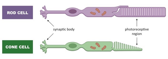 Image:
Cell Morphology
Retina Micrographs
Image:
Cell Morphology
Retina Micrographs
![]()
Understanding:
• Rods and cones differ in their sensitivities to light intensities and wavelengths
Rod Cells
- Rod cells function better in low light conditions (twilight vision) – they become quickly bleached in bright light
- Rod cells all contain the same pigment (rhodopsin) which absorbs a wide range of wavelengths
- Rod cells cannot differentiate between different colours (monochromatic)
- Rod cells are abundant at the periphery of the retina and hence are responsible for peripheral vision
- Rod cells produce poorly resolved images as many rod cells synapse with a single bipolar neuron
Cone Cells
- Cone cells function better in bright light conditions (daylight vision) – they require more photons of light to become activated
- There are three different types of cone cells, each with a different pigment that absorbs a narrow range of wavelengths
- Cone cells can therefore differentiate between different colours (red, blue and green)
- Cone cells are abundant at the centre of the retina (within the fovea) and hence are involved in visual focusing
- Cone cells produce well defined images as each cone cell synapses with a single bipolar neuron
Comparison of Rods and Cones

![]()
Understanding:
• Bipolar cells send the impulses from rods and cones to ganglion cells
• Ganglion cells send messages to the brain via the optic nerve
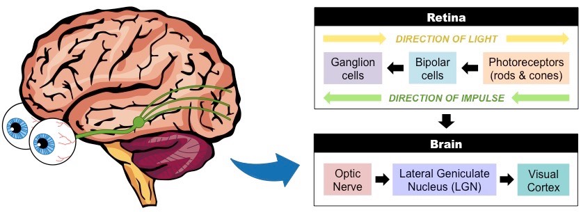
Photoreceptors (rods and cones) convert light stimuli into an electrical nerve impulse (action potential)
- This neural information is relayed to the brain via bipolar cells and ganglion cells
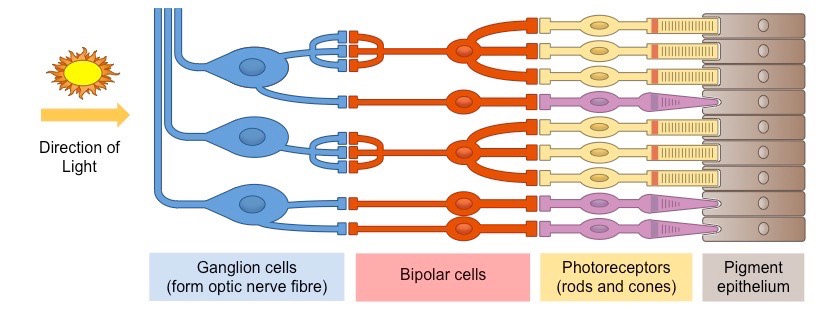
Bipolar cells transmit the nerve impulses produced by the photoreceptors to ganglion cells
- Many rod cells may synapse with a single bipolar cell, resulting in low resolution of sensory information (poor acuity)
- Most cone cells only synapse with a single bipolar cell, resulting in high resolution of sensory information (high acuity)
Ganglion cells transmit nerve impulses to the brain via long axonal fibres that compose the optic nerve
- Signals from ganglion cells may be sent to the visual cortex to form a composite representation of surroundings (i.e. sight)
- Alternatively, signals may be sent to other brain regions to coordinate eye movements or maintain circadian rhythms
There are no photoreceptors present in the region of the retina where ganglion axon fibres feed into the optic nerve
- This region is called the 'blind spot’ as visual information cannot be processed at this location
- The brain interpolates details from the surrounding regions, such that individuals do not perceive a visual blind spot
Transmission of Visual Stimuli
⇒ Click on the diagram to review the structure of the retina