![]()
Understanding:
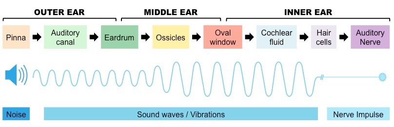
• Impulses caused by sound perception are transmitted to the brain via the auditory nerve
Sound travels as pressure waves in the air, which travel down the auditory canal and cause the eardrum to vibrate
- The degree of vibration of the eardrum (tympanic membrane) will depend on the frequency and amplitude of the sound wave
The eardrum transfers the vibrations via the bones of the middle ear (the ossicles) to the oval window of the cochlea
- The function of these bones is to amplify the vibrations from the eardrum (can increase magnification by ~ 20 times)
The vibration of the oval window causes fluid within the cochlea to be displaced – this displacement is detected by hair cells
- Activation of these hair cells generates nerve impulses which are transmitted via the auditory nerve to the brain
Overview of Sound Perception
![]()
Understanding:
• Structures in the middle ear transmit and amplify sound
The middle ear is separated from the outer ear by the eardrum and the inner ear by the oval window
- It is an air-filled chamber that houses three small bones (collectively called the ossicles)
The bones of the middle ear are individually called the malleus (hammer), incus (anvil) and stapes (stirrup)
- The malleus is in contact with the eardrum and the stapes contacts the oval window (while the incus connects the two)
The function of the ossicles is to amplify the sound vibrations by acting like levers to reduce the force distribution
- Sound travelling through air is mostly reflected when contacted by a liquid medium (due to the incompressibility of fluids)
- The amplification of sound by the ossicles allows the vibrational pressure to pass to the cochlear fluid with very little loss
- The oval window is smaller than the ear drum, which also assists in amplifying the sound energy
Sound Amplification by the Ossicles
![]()
Understanding:
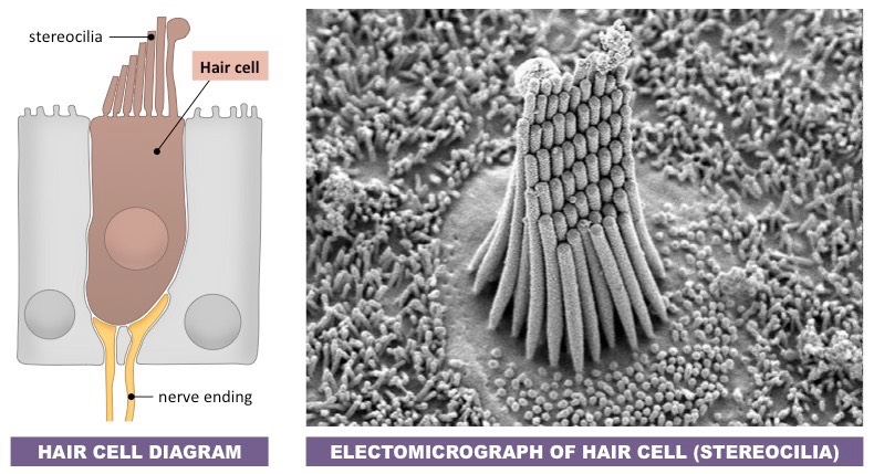
• Sensory hairs of the cochlea detect sounds at specific wavelengths
The cochlea is a fluid-filled spiral tube within the inner ear that converts sound vibrations into nerve impulses
- Displacement of fluid by sound vibrations activates sensory hair cells within the spiral part of the cochlea (organ of Corti)
Hair cells are mechanoreceptors that possess tiny hair-like extensions called stereocilia
- The cilia on hair cells vary in length and will each resonate to a different frequency of sound (i.e. specific wavelengths)
When the stereocilia are moved by the cochlear fluid, the hair cell will depolarise to generate a nerve impulse
- The nerve impulse will be transmitted via the auditory nerve to the auditory centres of the brain
- The kinetic movement of the cochlear fluid (and stereocilia motion) is dissipated by the vibration of the round window
Sensory Hair Cells