
![]()
Skill:
• Identification of tissue layers in transverse sections of the small intestine viewed with a microscope or in a
micrograph
The human intestines function to absorb the products of digestion and have specialised structures to fulfil this function
- The small intestine absorbs usable food substances (i.e. nutrients – monosaccharides, amino acids, fatty acids, vitamins, etc.)
- The large intestine absorbs water and dissolved minerals (i.e. ions) from the indigestible food residues
Structure of the Small Intestine
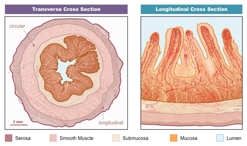
The small intestine is composed of four main tissue layers, which are (from outside to centre):
- Serosa – a protective outer covering composed of a layer of cells reinforced by fibrous connective tissue
- Muscle layer – outer layer of longitudinal muscle (peristalsis) and inner layer of circular muscle (segmentation)
- Submucosa – composed of connective tissue separating the muscle layer from the innermost mucosa
- Mucosa – a highly folded inner layer which absorbs material through its surface epithelium from the intestinal lumen
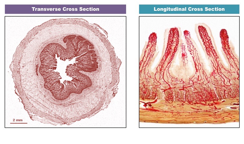
Below is a cross-section of the ileum – the final section of the small intestine (click on image to view coloured layers):
![]()
Understanding:
• Villi increase the surface area of epithelium over which absorption is carried out
• Villi absorb monomers formed by digestion as well as mineral ions and vitamins
The inner epithelial lining of the intestine is highly folded into finger-like projections called villi (singular: villus)
- Many villi will protrude into the intestinal lumen, greatly increasing the available surface area for material absorption
Features of Villi
Intestinal villi contain several key features which facilitate the absorption of digestive products (monomers, ions and vitamins):
- Microvilli – Ruffling of epithelial membrane further increases surface area
- Rich blood supply – Dense capillary network rapidly transports absorbed products
- Single layer epithelium – Minimises diffusion distance between lumen and blood
- Lacteals – Absorbs lipids from the intestine into the lymphatic system
- Intestinal glands – Exocrine pits (crypts of Lieberkuhn) release digestive juices
- Membrane proteins – Facilitates transport of digested materials into epithelial cells
Mnemonic: MR SLIM
Features of Intestinal Villi

Structure of Villus Epithelium
The epithelial lining of villi contains several structural features which optimise its capacity to absorb digested materials:
Tight Junctions
- Occluding associations between the plasma membrane of two adjacent cells, creating an impermeable barrier
- They keep digestive fluids separated from tissues and maintain a concentration gradient by ensuring one-way movement
Microvilli
- Microvilli borders significantly increase surface area of the plasma membrane (>100×), allowing for more absorption to occur
- The membrane will be embedded with immobilised digestive enzymes and channel proteins to assist in material uptake
Mitochondria
- Epithelial cells of intestinal villi will possess large numbers of mitochondria to provide ATP for active transport mechanisms
- ATP may be required for primary active transport (against gradient), secondary active transport (co-transport) or pinocytosis
Pinocytotic Vesicles
- Pinocytosis (‘cell-drinking’) is the non-specific uptake of fluids and dissolved solutes (a quick way to translocate in bulk)
- These materials will be ingested via the breaking and reforming of the membrane and hence contained within a vesicle
Cross-Section of Villus Epithelium
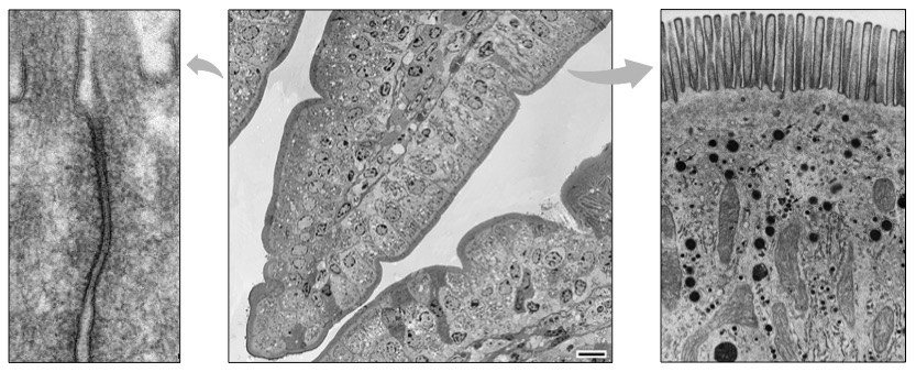
Structure: Pinocytotic Vesicles Tight Junction Microvilli Mitochondria All