
![]()
Skill:
• Drawing of the ultrastructure of prokaryotic cells based on electron micrographs
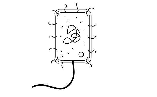
Prokaryotic Cell (click to label)
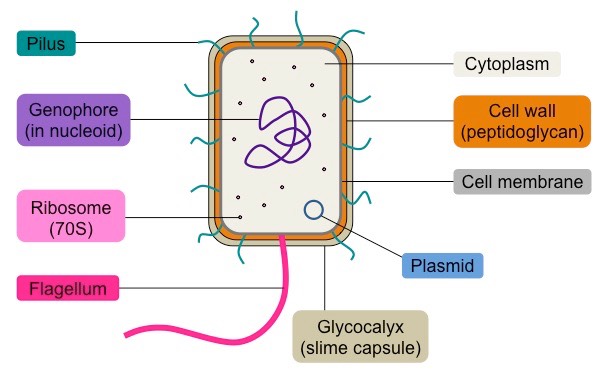
Key Features:
- Pili – shown as single lines
- Flagella – shown as thicker and significantly longer lines than the pili
- Ribosomes – labelled as 70S
- Cell wall – labelled as being composed of peptidoglycan; thicker than cell membrane
- Shape – appropriate for bacteria chosen (e.g. E. coli is a rod-shaped bacillus)
- Size – appropriate dimensions (e.g. length of cell twice the width)