
![]()
Application:
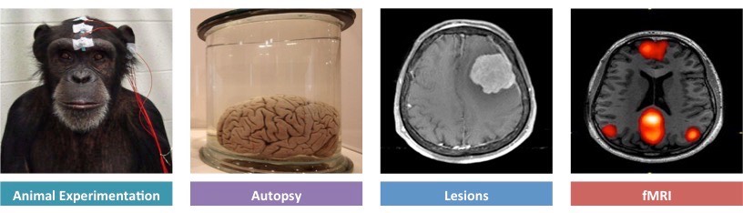
• Use of animal experiments, autopsy, lesions and fMRI to identify the role of different brain parts
The role of a specific brain part can be identified by either stimulating or removing the region to assess its effect
- Identification of brain roles can be made via the use of animal experiments, autopsy, lesions and fMRI
Animal Experiments
- Animal experimentation can be used to identify function by stimulating regions with electrodes or removing via lobotomy
- Because such methods are highly invasive and potentially damaging, animal models are frequently used
- Experimentation on animals involves less ethical restrictions than human studies (although ethical standards do exist)
- Animal studies are limited by the differences between animal and human brains, making valid comparisons difficult
- Example: Animal studies using mice and rats have been used to develop drug treatments for diseases such as MS
Lesions
- Lesions are abnormal areas of brain tissue which can indicate the effect of the loss of a brain area
- Lesions can be identified via post-mortem analysis (autopsy) or via scans of the brain (CT scans or MRI)
- The effects of lesions can be difficult to identify, as many functions may involve multiple brain areas
- Additionally, the brain has the capacity to re-learn certain skills by re-routing instructions to other areas (plasticity)
- Example: Split brain patients have been used to identify specific roles of the left and right cerebral hemisphere
Autopsy
- An autopsy is a post-mortem examination of a corpse via dissection in order to evaluate causes of death
- Comparisons can be made between the brains of healthy and diseased corpses to identify affected brain areas
- Example: Cadavers who suffered from aphasia (language impairment) in life demonstrate damage to specific areas
fMRI
- Functional magnetic resonance imaging (fMRI) records changes in blood flow within the brain to identify activated areas
- Oxygenated haemoglobin responds differently to a magnetic field than deoxygenated haemoglobin
- These differences in oxygenation can be represented visually and reflect differences in the level of brain activity
- fMRI is non-invasive and can be used to identify multiple brain regions involved in complex, integrated brain activities
- Example: fMRI studies have been used to diagnose ADHD and dyslexia, as well as monitor recovery from strokes
Methods for Identifying Brain Functions
![]()
Application:
• Visual cortex, Broca’s area, nucleus accumbens as areas of the brain with specific functions
While complex activities may require integration of multiple regions, some specific functions are localised to particular areas
- Examples of brain areas with clearly defined functions include the visual cortex, Broca’s area and the nucleus accumbens
Visual Cortex
- Located within the occipital lobe of the cerebrum and receives neural impulses from light-sensitive cells in the eyes
- The visual cortex is the region of the brain responsible for visual perception (sight)
Broca’s Area
- Located within the frontal lobe of the left cerebral hemisphere (not present in the right hemisphere)
- Is responsible for speech production (if damaged, the individual cannot produce meaningful speech despite intending to)
Nucleus Accumbens
- The nucleus accumbens is involved in the pleasure reward pathway and is found within each cerebral hemisphere
- It secretes neurotransmitters responsible for feelings of pleasure (dopamine) and satiety (serotonin)
- It communicates with other centres involved in the mechanisms of pleasure, such as the ventral tegmental area (VTA)